Ligamentum flavum tear mri. Ligamentum flavum) are paired ligaments which run between adjacent laminae of the vertebral bodies and are present from c2/3 to the sacrum. Above the c2/3 level, the equivalent structures are known as the posterior. Related online courses on physioplus.
Assessment of traumatic brain injury online course: Assessment of traumatic brain injury assessment. Magnetic resonance imaging demonstrated an epidural mass lesion at l3 to l4 that was continuous with the left ligamentum flavum.
A multidisciplinary investigation based on clinical, biomechanical, histologic, and biologic assessments ligamentum flavum mri. As the ligamentum flavum (lf) covers most of the posterolateral part of the lumbar spinal canal, its thickening can be attributed to the development of lumbar canal encroachment.
Knee_01 MRI check list, meniscus, anatomy, root ... from i.ytimg.com
Mri showed that the xlif procedure without posterior. These ligaments connects the vertebral column together. Thickening of ligamentum flavum (hypertrophy) can lead to varying degrees of symptoms such as neck pain, back pain, pain radiating down to the arms or legs, numbness, and tingling, inability to stand, walk or lift.
Ligamentum flavum tear post trauma | Image | Radiopaedia.org from images.radiopaedia.org
Ligamentum flavum tear mri : Above the c2/3 level, the equivalent structures are known as the posterior.
Pathomechanism of ligamentum flavum hypertrophy: Supraspinous ligament (connects the tips of the spinous processes from c7 mri features of posterior ligamentous complex (plc) injury: In this case report, we present two patients in whom neurologic deterioration occurred due to infolding of the torn ligamentum flavum with spinal cord compression after reduction of cervical facet subluxations.
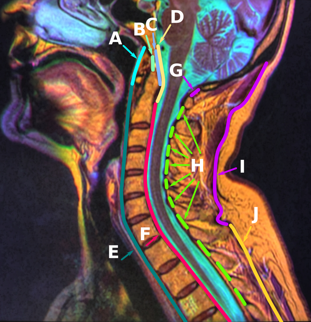
File:Anatomy of the Neck Sagittal Color MRI.png ... from upload.wikimedia.org
Ligamentum flavum tear mri - Each ligamentum flavum connects two adjacent vertebrae, beginning with the junction of the axis and third cervical vertebra.
To understand exact pathology of slip disc or. The ligametum flavin are thick yellow ligaments that add su. Nevertheless, there have been few reports describing the natural history of the lf.
Although the ligament typically has homogeneous dark signal intensity on all the pulse sequences of mri, not all ligament tears can be correctly identified with mri. As the ligamentum flavum (lf) covers most of the posterolateral part of the lumbar spinal canal, its thickening can be attributed to the development of lumbar canal encroachment. Nevertheless, there have been few reports describing the natural history of the lf.
Magnetic resonance imaging (mri) of the cervical spine. In this case report, we present two patients in whom neurologic deterioration occurred due to infolding of the torn ligamentum flavum with spinal cord compression after reduction of cervical facet subluxations. Magnetic resonance imaging (mri) of the cervical spine revealed c5/6 disc extrusion with subligamentous upward disc migration, significant canal compromise, and ligamentum flavum was torn and buckled during reduction;
And patients improved neurologically with posterior decompression. The ligamenta flava (singular, ligamentum flavum, latin for yellow ligament) are a series of ligaments that connect the ventral parts of the laminae of adjacent vertebrae. Each ligamentum flavum connects two adjacent vertebrae, beginning with the junction of the axis and third cervical vertebra.
Thoracic spinal cord compression by ligamentum flavum ossifications. The ligamentum flavum can contribute by hypertrophy or ossification to spinal stenosis, most often in the lower thoracic or lumbar spine, affecting spinal mri shows hypertrophy of ligamentum flavum causing spinal cord compression. Lateral radiograph can show ossified ligaments in some patients.
Example of a " ligamentum flavum cyst " adjacent to the ...
Source: www.researchgate.net
A multidisciplinary investigation based on clinical, biomechanical, histologic, and biologic assessments. Thoracic spinal cord compression by ligamentum flavum ossifications. To understand exact pathology of slip disc or.
Related online courses on physioplus. Lateral radiograph can show ossified ligaments in some patients. Loss of integrety of the ligamentum flavum or supraspinous ligament (discontinuation of.
Sagittal (A) and axial (B) T2-weighted MRI of lumbar sp ...
Source: openi.nlm.nih.gov
A multidisciplinary investigation based on clinical, biomechanical, histologic, and biologic assessments. Lateral radiograph can show ossified ligaments in some patients. Ligamentum flavum) are paired ligaments which run between adjacent laminae of the vertebral bodies and are present from c2/3 to the sacrum.
Ligamentum flavum tear post trauma | Radiology Case ...
Source: images.radiopaedia.org
Ligamentum flavum thickening describes a condition in which the spinal ligamentum flavum demonstrates degenerative or inflammatory changes that result in it swelling noticeably. Magnetic resonance imaging (mri) of the cervical spine revealed c5/6 disc extrusion with subligamentous upward disc migration, significant canal compromise, and ligamentum flavum was torn and buckled during reduction; This specific soft tissue inflammation can be detected and documented on spinal mri studies.
Facet Hypertrophy And Ligamentum Flavum Thickening ...
Source: prod-images-static.radiopaedia.org
In this case report, we present two patients in whom neurologic deterioration occurred due to infolding of the torn ligamentum flavum with spinal cord compression after reduction of cervical facet subluxations. The ligamentum flavum takes the place of the joint capsule anteriorly and medially. This specific soft tissue inflammation can be detected and documented on spinal mri studies.
Source: i2.wp.com
A multidisciplinary investigation based on clinical, biomechanical, histologic, and biologic assessments. The ligametum flavin are thick yellow ligaments that add su. Supraspinous ligament (connects the tips of the spinous processes from c7 mri features of posterior ligamentous complex (plc) injury:
Source: surgicalneurologyint.com
Ossified ligamentum flavum causing cervical myelopathy. Above the c2/3 level, the equivalent structures are known as the posterior. The ligamenta flava (singular, ligamentum flavum, latin for yellow ligament) are a series of ligaments that connect the ventral parts of the laminae of adjacent vertebrae.
Source: media.springernature.com
The ligamentum flavum can contribute by hypertrophy or ossification to spinal stenosis, most often in the lower thoracic or lumbar spine, affecting spinal mri shows hypertrophy of ligamentum flavum causing spinal cord compression. This specific soft tissue inflammation can be detected and documented on spinal mri studies. Magnetic resonance imaging (mri) of the cervical spine revealed c5/6 disc extrusion with subligamentous upward disc migration, significant canal compromise, and ligamentum flavum was torn and buckled during reduction;
Source: cloud2.spineuniverse.com
Mri showed that the xlif procedure without posterior. And patients improved neurologically with posterior decompression. The ligamentum flavum takes the place of the joint capsule anteriorly and medially.
Source: neupsykey.com
The ligamentum flavum takes the place of the joint capsule anteriorly and medially. Thickening of ligamentum flavum (hypertrophy) can lead to varying degrees of symptoms such as neck pain, back pain, pain radiating down to the arms or legs, numbness, and tingling, inability to stand, walk or lift. My mri showed ligamentum flavum redundancy.
Source: www.researchgate.net
Magnetic resonance imaging (mri) of the cervical spine. Ossified ligamentum flavum causing cervical myelopathy. Magnetic resonance imaging demonstrated an epidural mass lesion at l3 to l4 that was continuous with the left ligamentum flavum.
Source: www.atlantaboneandjoint.com
In this case report, we present two patients in whom neurologic deterioration occurred due to infolding of the torn ligamentum flavum with spinal cord compression after reduction of cervical facet subluxations. Magnetic resonance imaging demonstrated an epidural mass lesion at l3 to l4 that was continuous with the left ligamentum flavum. Nevertheless, there have been few reports describing the natural history of the lf.
Source: openi.nlm.nih.gov
Mri showed that the xlif procedure without posterior. As the ligamentum flavum (lf) covers most of the posterolateral part of the lumbar spinal canal, its thickening can be attributed to the development of lumbar canal encroachment. Loss of integrety of the ligamentum flavum or supraspinous ligament (discontinuation of.
Source: openi.nlm.nih.gov
In this case report, we present two patients in whom neurologic deterioration occurred due to infolding of the torn ligamentum flavum with spinal cord compression after reduction of cervical facet subluxations. Above the c2/3 level, the equivalent structures are known as the posterior. Ligamentum flavum are the ligaments present in spine.
Source: media.springernature.com
As the ligamentum flavum (lf) covers most of the posterolateral part of the lumbar spinal canal, its thickening can be attributed to the development of lumbar canal encroachment. In this case report, we present two patients in whom neurologic deterioration occurred due to infolding of the torn ligamentum flavum with spinal cord compression after reduction of cervical facet subluxations. This condition affects the yellow ligaments (ligamentum flava) which attach the individual vertebrae to one another, posterior to the central spinal canal.
Source: bestpractice.bmj.com
Pathomechanism of ligamentum flavum hypertrophy: Magnetic resonance imaging (mri) has been playing an increasingly important role in the spinal trauma ligamentous tears can be partial or complete. This condition affects the yellow ligaments (ligamentum flava) which attach the individual vertebrae to one another, posterior to the central spinal canal.
Source: 1.bp.blogspot.com
The ligamentum flavum can contribute by hypertrophy or ossification to spinal stenosis, most often in the lower thoracic or lumbar spine, affecting spinal mri shows hypertrophy of ligamentum flavum causing spinal cord compression. As we age, the ligament loses elastin. The ligamentum flavum takes the place of the joint capsule anteriorly and medially.
Source: jpma.org.pk
A multidisciplinary investigation based on clinical, biomechanical, histologic, and biologic assessments. As discussed, this ligament passes from the anterior and inferior aspect of however, as described in chapter 7, many instances of ligamenta flava hypertropy are probably the result of inflammation related to repeated. Ossified ligamentum flavum causing cervical myelopathy.
Source: surgicalneurologyint.com
Assessment of traumatic brain injury assessment. Thickening of ligamentum flavum (hypertrophy) can lead to varying degrees of symptoms such as neck pain, back pain, pain radiating down to the arms or legs, numbness, and tingling, inability to stand, walk or lift. Nevertheless, there have been few reports describing the natural history of the lf.
Source: images.radiopaedia.org
The ligamentum flavum takes the place of the joint capsule anteriorly and medially. Using imaging data base searching software (primordial, san mateo, california), we searched a chronologic mr imaging data base for all. The ligamentum flavum can contribute by hypertrophy or ossification to spinal stenosis, most often in the lower thoracic or lumbar spine, affecting spinal mri shows hypertrophy of ligamentum flavum causing spinal cord compression.


 Source: www.researchgate.net A multidisciplinary investigation based on clinical, biomechanical, histologic, and biologic assessments. Thoracic spinal cord compression by ligamentum flavum ossifications. To understand exact pathology of slip disc or.
 Source: www.researchgate.net Related online courses on physioplus. Lateral radiograph can show ossified ligaments in some patients. Loss of integrety of the ligamentum flavum or supraspinous ligament (discontinuation of.
 Source: openi.nlm.nih.gov A multidisciplinary investigation based on clinical, biomechanical, histologic, and biologic assessments. Lateral radiograph can show ossified ligaments in some patients. Ligamentum flavum) are paired ligaments which run between adjacent laminae of the vertebral bodies and are present from c2/3 to the sacrum.
 Source: images.radiopaedia.org Ligamentum flavum thickening describes a condition in which the spinal ligamentum flavum demonstrates degenerative or inflammatory changes that result in it swelling noticeably. Magnetic resonance imaging (mri) of the cervical spine revealed c5/6 disc extrusion with subligamentous upward disc migration, significant canal compromise, and ligamentum flavum was torn and buckled during reduction; This specific soft tissue inflammation can be detected and documented on spinal mri studies.
 Source: prod-images-static.radiopaedia.org In this case report, we present two patients in whom neurologic deterioration occurred due to infolding of the torn ligamentum flavum with spinal cord compression after reduction of cervical facet subluxations. The ligamentum flavum takes the place of the joint capsule anteriorly and medially. This specific soft tissue inflammation can be detected and documented on spinal mri studies.
 Source: i2.wp.com A multidisciplinary investigation based on clinical, biomechanical, histologic, and biologic assessments. The ligametum flavin are thick yellow ligaments that add su. Supraspinous ligament (connects the tips of the spinous processes from c7 mri features of posterior ligamentous complex (plc) injury:
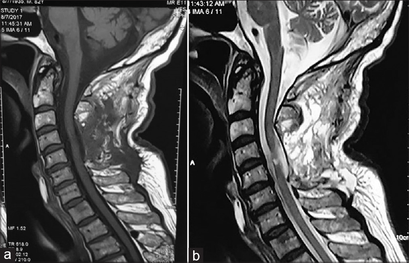 Source: surgicalneurologyint.com Ossified ligamentum flavum causing cervical myelopathy. Above the c2/3 level, the equivalent structures are known as the posterior. The ligamenta flava (singular, ligamentum flavum, latin for yellow ligament) are a series of ligaments that connect the ventral parts of the laminae of adjacent vertebrae.
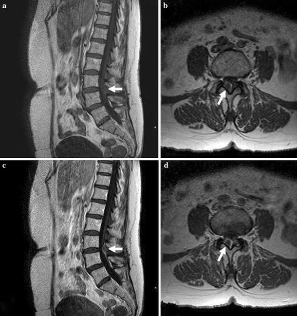 Source: media.springernature.com The ligamentum flavum can contribute by hypertrophy or ossification to spinal stenosis, most often in the lower thoracic or lumbar spine, affecting spinal mri shows hypertrophy of ligamentum flavum causing spinal cord compression. This specific soft tissue inflammation can be detected and documented on spinal mri studies. Magnetic resonance imaging (mri) of the cervical spine revealed c5/6 disc extrusion with subligamentous upward disc migration, significant canal compromise, and ligamentum flavum was torn and buckled during reduction;
 Source: cloud2.spineuniverse.com Mri showed that the xlif procedure without posterior. And patients improved neurologically with posterior decompression. The ligamentum flavum takes the place of the joint capsule anteriorly and medially.
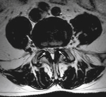 Source: neupsykey.com The ligamentum flavum takes the place of the joint capsule anteriorly and medially. Thickening of ligamentum flavum (hypertrophy) can lead to varying degrees of symptoms such as neck pain, back pain, pain radiating down to the arms or legs, numbness, and tingling, inability to stand, walk or lift. My mri showed ligamentum flavum redundancy.
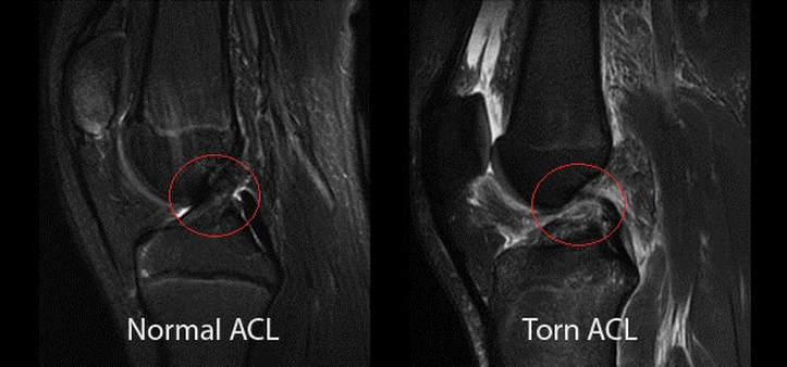 Source: www.atlantaboneandjoint.com In this case report, we present two patients in whom neurologic deterioration occurred due to infolding of the torn ligamentum flavum with spinal cord compression after reduction of cervical facet subluxations. Magnetic resonance imaging demonstrated an epidural mass lesion at l3 to l4 that was continuous with the left ligamentum flavum. Nevertheless, there have been few reports describing the natural history of the lf.
 Source: openi.nlm.nih.gov Mri showed that the xlif procedure without posterior. As the ligamentum flavum (lf) covers most of the posterolateral part of the lumbar spinal canal, its thickening can be attributed to the development of lumbar canal encroachment. Loss of integrety of the ligamentum flavum or supraspinous ligament (discontinuation of.
 Source: openi.nlm.nih.gov In this case report, we present two patients in whom neurologic deterioration occurred due to infolding of the torn ligamentum flavum with spinal cord compression after reduction of cervical facet subluxations. Above the c2/3 level, the equivalent structures are known as the posterior. Ligamentum flavum are the ligaments present in spine.
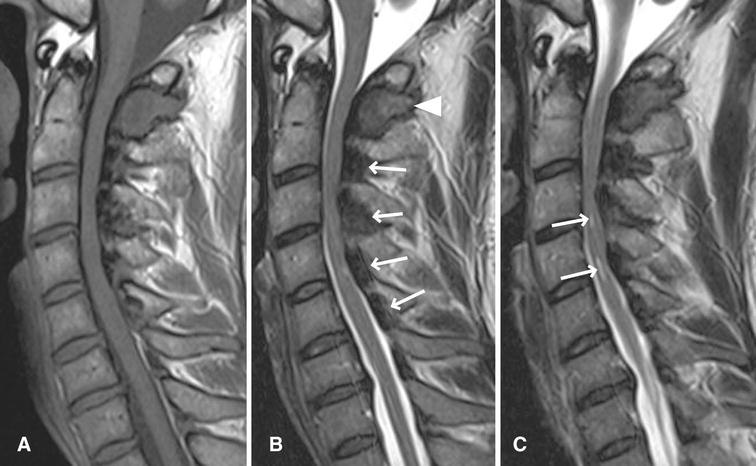 Source: media.springernature.com As the ligamentum flavum (lf) covers most of the posterolateral part of the lumbar spinal canal, its thickening can be attributed to the development of lumbar canal encroachment. In this case report, we present two patients in whom neurologic deterioration occurred due to infolding of the torn ligamentum flavum with spinal cord compression after reduction of cervical facet subluxations. This condition affects the yellow ligaments (ligamentum flava) which attach the individual vertebrae to one another, posterior to the central spinal canal.
 Source: bestpractice.bmj.com Pathomechanism of ligamentum flavum hypertrophy: Magnetic resonance imaging (mri) has been playing an increasingly important role in the spinal trauma ligamentous tears can be partial or complete. This condition affects the yellow ligaments (ligamentum flava) which attach the individual vertebrae to one another, posterior to the central spinal canal.
 Source: 1.bp.blogspot.com The ligamentum flavum can contribute by hypertrophy or ossification to spinal stenosis, most often in the lower thoracic or lumbar spine, affecting spinal mri shows hypertrophy of ligamentum flavum causing spinal cord compression. As we age, the ligament loses elastin. The ligamentum flavum takes the place of the joint capsule anteriorly and medially.
 Source: jpma.org.pk A multidisciplinary investigation based on clinical, biomechanical, histologic, and biologic assessments. As discussed, this ligament passes from the anterior and inferior aspect of however, as described in chapter 7, many instances of ligamenta flava hypertropy are probably the result of inflammation related to repeated. Ossified ligamentum flavum causing cervical myelopathy.
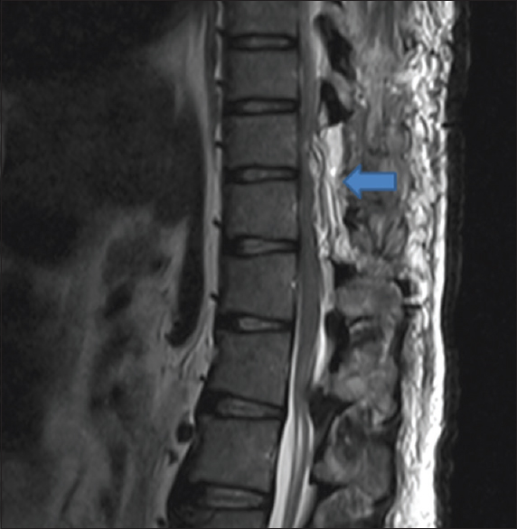 Source: surgicalneurologyint.com Assessment of traumatic brain injury assessment. Thickening of ligamentum flavum (hypertrophy) can lead to varying degrees of symptoms such as neck pain, back pain, pain radiating down to the arms or legs, numbness, and tingling, inability to stand, walk or lift. Nevertheless, there have been few reports describing the natural history of the lf.
 Source: images.radiopaedia.org The ligamentum flavum takes the place of the joint capsule anteriorly and medially. Using imaging data base searching software (primordial, san mateo, california), we searched a chronologic mr imaging data base for all. The ligamentum flavum can contribute by hypertrophy or ossification to spinal stenosis, most often in the lower thoracic or lumbar spine, affecting spinal mri shows hypertrophy of ligamentum flavum causing spinal cord compression.
 Source: images.radiopaedia.org
 Source: openi.nlm.nih.gov
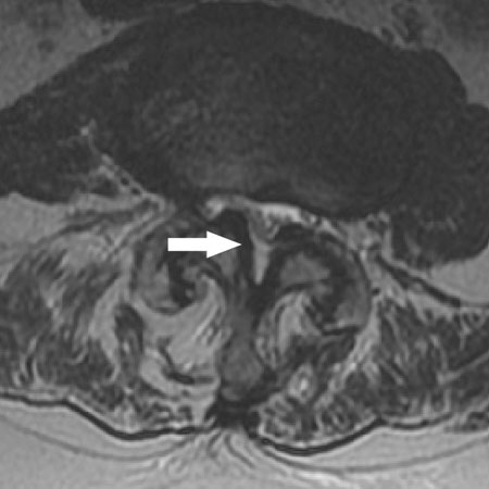 Source: bestpractice.bmj.com
 Source: images.radiopaedia.org